OptiFlow is a domain-aware image-analytics platform for life-science imaging — from light, fluorescence and confocal microscopy to super-resolution and whole-slide pathology. Build the analysis visually, quantify what you see, and run it from a single image to a whole cohort — exporting results that carry the exact method that made them. No code, 100% local.
Life-science imaging has outpaced the tools used to analyse it. Methods grow so intricate they no longer fit in a paragraph of a methods section; biologists are pushed into scripting they never signed up for; and the volumes from high-content screens and whole-slide scanners are far beyond what anyone can review by hand.
A multi-step analysis rarely survives the journey from one operator, instrument, or paper to the next — and a result you can't reproduce is a result you can't defend.
Researchers lose experimental time to image processing and scripting instead of the biology they were hired for — and pipelines break the moment the person who built them moves on.
Whole-slide and high-content datasets are too large to inspect exhaustively, yet too important to sample — so the rare, decisive event is the one most easily missed.
Biomedical imaging spans very different teams with very different goals — from a single time-point to longitudinal, live-cell studies — but the same need for analysis they can trust, repeat, and defend. OptiFlow adapts to each, rather than forcing your work into a generic mould.
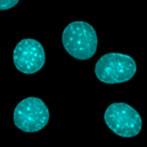
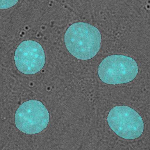
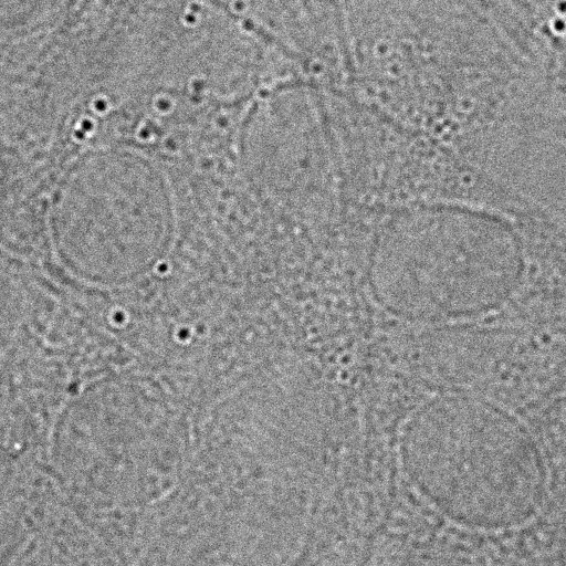
OptiFlow's CytoByte engine performs virtual nuclear staining directly from label-free DIC or phase contrast — in real time, with no chemistry and no phototoxicity, revealing native biology undisturbed. Because the cells are never perturbed, live and longitudinal experiments stay clean.
Virtual nuclei · CytoByte
DIC · label-free
Image live cells as often as you like, without the photodamage a fluorescent stain introduces.
No dyes, no incubation, no fixation — skip the staining workflow and its cost and variability entirely.
Image the sample as it is, take your readout, and return it straight to the incubator — nothing is stained, fixed, or consumed.
The science behind CytoByte is peer-reviewed and proven in the field — not a marketing claim.
Stream the virtual staining live as the microscope acquires each frame — overlay the real and virtual channels for an AR view of your structure of interest.
Compose exactly the pipeline your assay needs from a deep library of domain-aware operations — purpose-built for quantitative microscopy. Preview any of them on your own images, then license only what your work demands.
Denoising that is classical, AI-guided, and hybrid — NLM, bilateral, wavelet, total-variation and BM3D for low-light live-cell and EM data. Illumination and contrast correction, CLAHE, and frequency-domain filtering for clean, comparable inputs.
Otsu, multi-Otsu and adaptive thresholding; morphology and connected-component labelling; ridge filters (Frangi, Sato, Meijering) for vessels, neurites and filaments. Calibrated area, distance and interactive line-intensity profiles — in real units, read from your metadata.
Track objects across frames and through z — motility, migration, division and lineage — turning live-cell time-lapse and 3D series into quantitative kinetic and spatiotemporal readouts.
Supervised deep learning for cell type, phenotype, or disease grade — trained on your own annotated images, so the model learns your problem rather than approximating it.
Exhaustive, slide-wide search for low-prevalence targets — circulating tumour cells, mitotic figures — plus unsupervised anomaly detection that flags the unexpected with no labels required.
Per-cell and population morphometrics, enumeration and texture analysis with batch normalisation built in — then built-in plotting (histogram, scatter, box-plot, violin, CDF) and automatic export to Excel and formatted PDF reports.
Channel and colour operations, lookup tables, geometry and alignment, type conversion, and pixel-wise image maths and compositing — the connective tissue that lets any workflow fit together exactly as your assay requires.
Modular by design — start with what today's work needs and add modules as your assays grow, instrument by instrument and lab by lab. Every operation runs locally, on your own machines. See the full OptiFlow platform.
Modern image analysis has become too complex to write into a methods section. OptiFlow records it for you — so every result is reproducible, every method is portable and submittable as-is, and your data never leaves your control.
The complete pipeline — every operation, parameter, calibration and operator — is written into the image itself, encrypted and readable only inside OptiFlow. An encrypted, rollback-resistant timestamp guards the record; where it matters, lock it to a password only your team holds.
Export the complete pipeline as a standalone file — to attach to a publication, hand to a collaborator, or release alongside open data. Anyone you give it to can reproduce the exact analysis, down to the parameter.
Processing reads and writes your own filesystem directly. No image data crosses the network — no cloud account, no exceptions — so patient data and unpublished results stay exactly where they belong.
Every result already contains the workflow that made it. So revisiting your own analysis, handing it to a collaborator, satisfying a reviewer, or re-running across a cohort needs nothing rebuilt and nothing re-documented — and the record stays encrypted, readable only inside OptiFlow, and accessible only when you choose.
Logic Byte's biomedical capabilities trace directly to peer-reviewed research and deployed systems — the foundation beneath every module on this page.
No rip-and-replace, no leap of faith. Prove the value on your own data first, then grow module by module, lab by lab.
Bring a biomedical imaging problem and your own data — we'll show you exactly what OptiFlow does with them. If your workflow needs something we haven't built yet, we'll build it.